

Whether an ADC succeeds or fails often turns on the dynamic balance of payload release. Premature release means a risk of systemic toxicity; insufficient release means compromised efficacy. A precise picture of how the payload behaves in the body is a threshold every ADC development team has to cross.
Following systematic validation, Sailybio is launching a series of anti-ADC payload monoclonal antibodies covering the five mainstream payload classes — DM1, DXd, MMAE, MMAF and SN38. The line gives ADC development programmes a full-process detection toolkit running from process quality control through to pharmacokinetic analysis.
1. ADC Drugs: How the Biological Missile Is Built
An ADC is built from three core components: a monoclonal antibody, a linker and a cytotoxic payload. The antibody works like a navigation system, recognising and locking onto antigens on the tumour cell surface. The linker governs the timing of toxin release. The payload — the warhead — is the decisive unit: once inside the tumour cell it destroys DNA or microtubule structures to achieve targeted elimination.
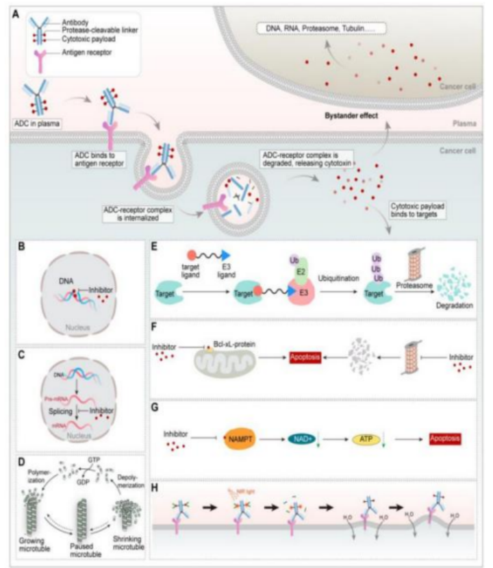 Figure 1. Structure and mechanism of action of antibody–drug conjugates.
2. Anti-Payload Antibodies: A Molecular Radar Aimed at the Toxin
Pharmacokinetic studies of an ADC need to cover in vitro stability, the plasma concentration–time curve, and absorption, distribution, metabolism and excretion. In practice three analytes are measured: conjugated antibody, total antibody, and free small-molecule compound.
Manufacturing process control: keeping every ADC batch consistent
In chemical conjugation, residual free toxin is a common source of batch-to-batch variation. Using a competitive ELISA, anti-payload antibodies detect residual free toxin efficiently. This helps manufacturers keep process quality under tight control, prevents batch rejection caused by excess free toxin, ensures every ADC batch meets specification, and lowers production risk.
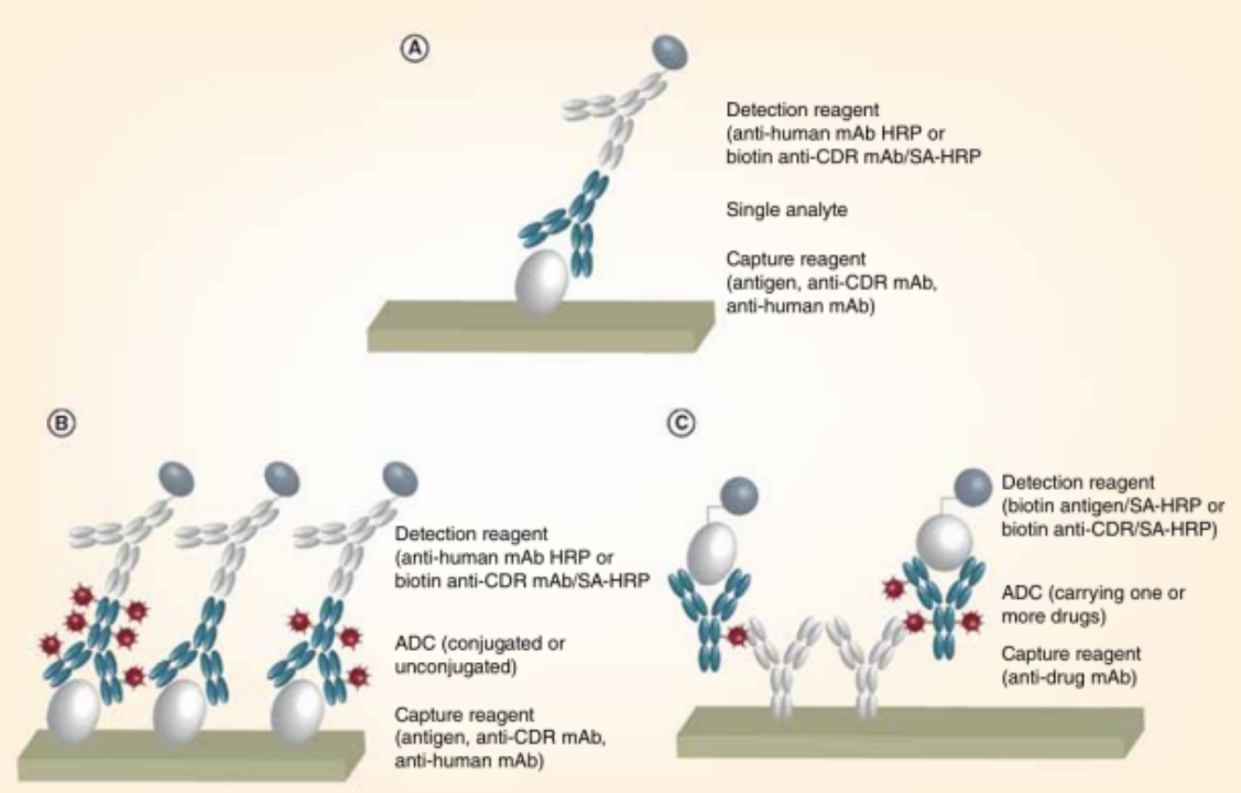
Figure 2. Ligand-binding assays. (A) Typical ELISA; (B) ADC total-antibody ELISA; (C) ADC conjugated-antibody ELISA.
Formulation stability studies: a scientific basis for formulation optimisation
The payload release rate of an ADC under different storage conditions — temperature, pH, light exposure — directly affects its shelf life and safety. Anti-payload antibodies monitor payload release in real time, helping R&D teams judge linker stability, providing the data to support formulation optimisation, extending shelf life and improving patient safety.
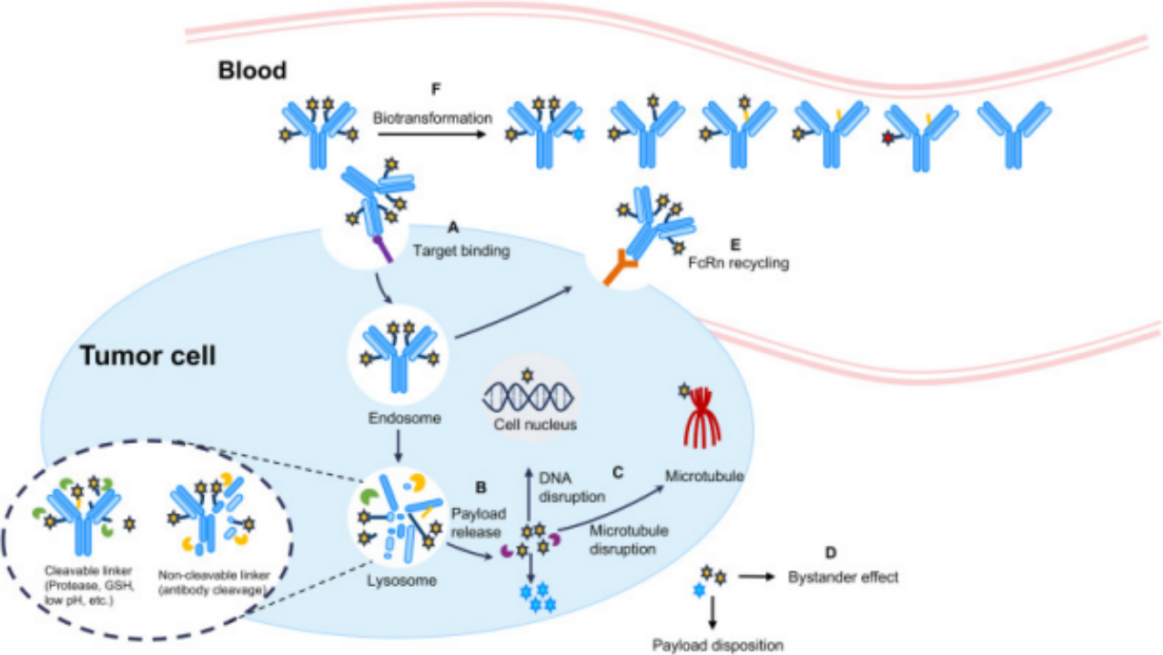
Figure 3. The biotransformation and disposition fate of typical ADCs following intravenous administration.
Pharmacokinetic analysis: one sample, three key readouts
Pharmacokinetics (PK) sits at the centre of ADC clinical filings. With anti-payload antibodies, researchers can establish an LC-MS/MS method that simultaneously quantifies intact ADC, total antibody and free toxin in plasma — three key quantitative readouts from a single workflow. This shortens the analytical cycle considerably, provides a reliable basis for dose design and toxicity assessment, and helps accelerate IND filings.
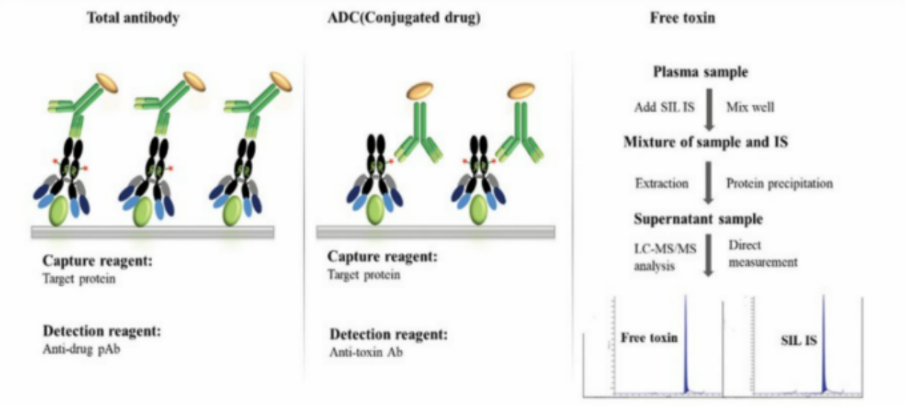
Figure 4. Common strategies for ADC bioanalysis.
Toxicity mechanism elucidation: driving the next iteration of linker technology
Off-target toxicity is one of the biggest challenges in ADC development. Using methods such as immunohistochemistry, anti-payload antibodies track payload distribution across tissues, showing which normal organs are under attack by free toxin and pinpointing the source of that toxicity. The evidence feeds back into linker design and optimisation, clearing the way for safer and more effective next-generation ADCs.
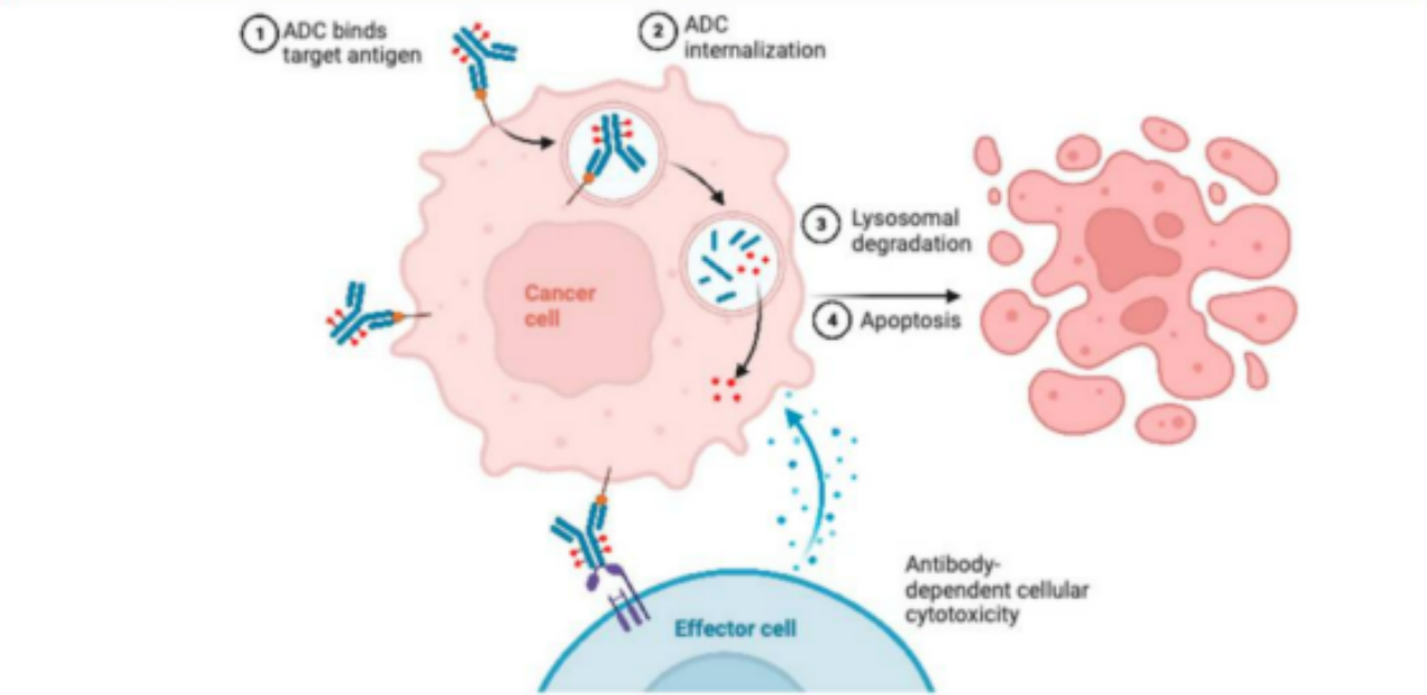 Figure 5. Possible resistance mechanisms based on ADC.
3. Anti-Payload Antibody Product Line
Built on a hybridoma technology platform, we have developed five anti-ADC payload monoclonal antibodies. All are mouse IgG1 isotype and are suitable for ELISA, sandwich ELISA and competitive ELISA formats.
Product Catalog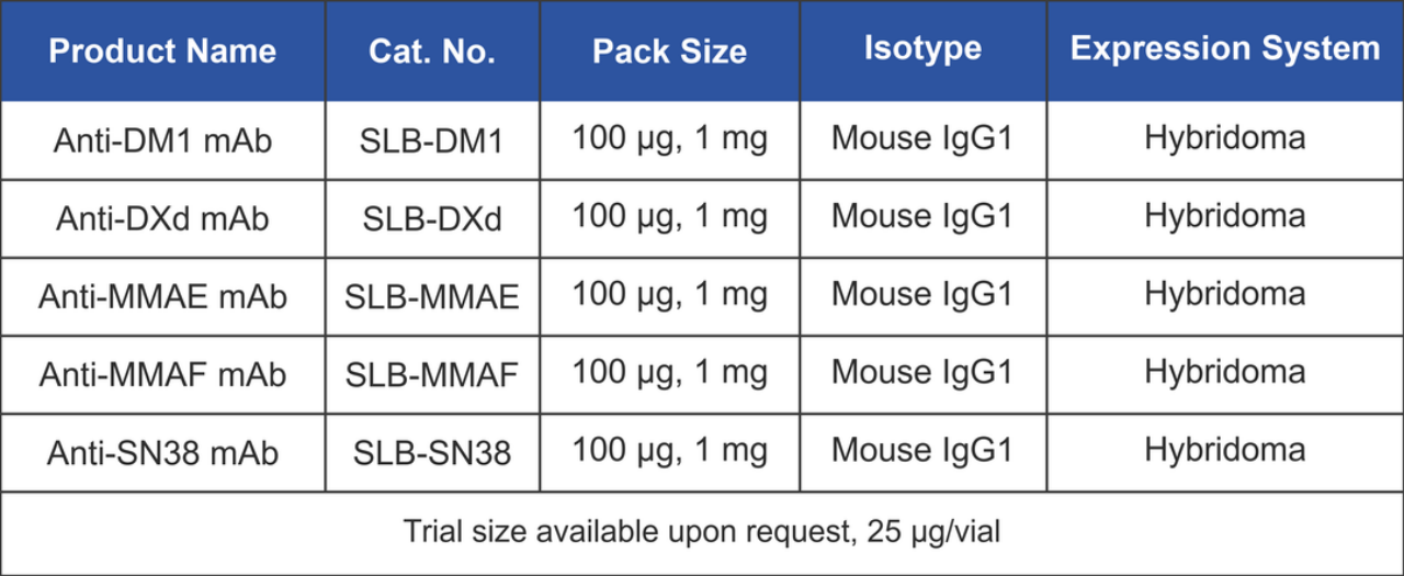 Table 1. Anti-ADC payload monoclonal antibody product line.
Product Data Examples
(1) Anti-DM1 mAb
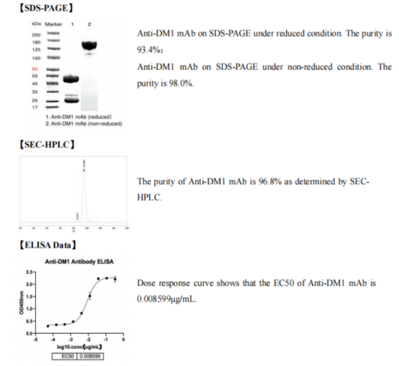
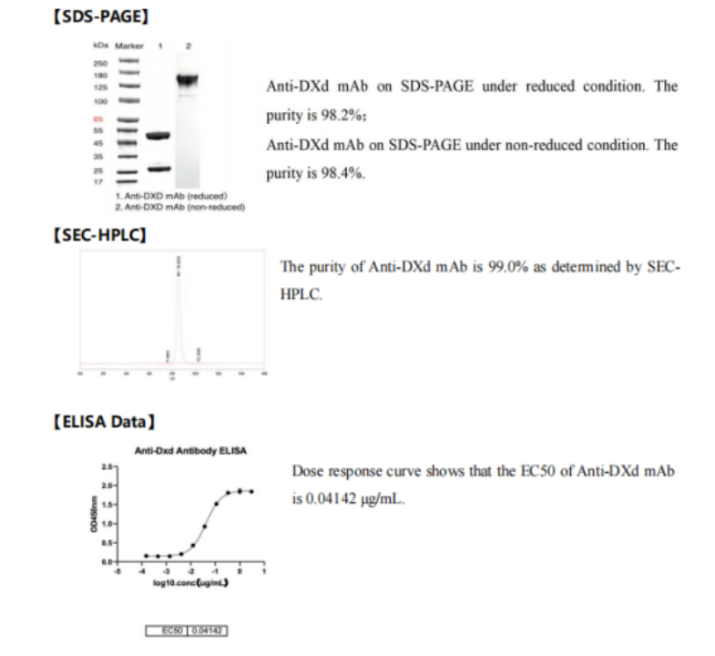
(2) Anti-DXd mAb